
Archivo:Pre- and post-central gyrus, right hemisphere.jpg

Tamaño de esta previsualización: 380 × 599 píxeles. Otras resoluciones: 152 × 240 píxeles · 304 × 480 píxeles · 884 × 1394 píxeles.
Ver la imagen en su resolución original (884 × 1394 píxeles; tamaño de archivo: 1,13 MB; tipo MIME: image/jpeg)
Resumen
| Descripción |
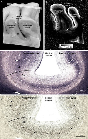
English: Pre- and post-central gyrus, right hemisphere. Microstructural 7-T MR mapping in post-mortem brains. (A) Tissue block (pre- and post-central gyrus, right hemisphere) from a post-mortem human brain (female, 61 years, died of pulmonary failure and chronic obstructive pulmonary disease, autopsy performed with informed consent of the patient’s relatives, post-mortem interval before fixation 24 h, fixed in 4% formalin for 2 months) prior to MR scanning and histological processing. M1 = primary motor cortex in the posterior wall of the precentral gyrus, S1 = primary somatosensory cortex in the anterior wall of the post-central gyrus. (B) Quantitative T1 map of the tissue block [for plane of sectioning see rectangle in (A)]. MP2RAGE sequence at 7 T, voxel size (0.33 mm)3, 32 averages, acquisition time 3 h 50 min, surrounding medium: Fomblin (Solvay Solexis, Bollate, Italy). Arrow indicates a sharp change in T1 contrast at the base of the precentral gyrus that matches a change in the myelo- and cytoarchitectonic pattern [cf. (C,D)]. (C,D) Frozen sections (30 μm) from a corresponding position of the same block stained for myelin basic protein [rat monoclonal antibody, avidin–biotin–peroxidase complex (ABC) method, chromogen: DAB and ammonium nickel(II) sulfate (C)] and cell bodies. Micrographs show the fundus of the central sulcus [same orientation as in (A,B)]. The drop in T1 values at the base of the precentral gyrus coincides with an increase in myelin basic protein immunostaining [line in (C)]. In an accompanying section stained for cell bodies, this position is characterized by an increase in gray matter thickness, a disappearing inner granular layer (asterisks), and emerging giant pyramidal (Betz) cells (arrowheads). This transition [lines in (C,D)] corresponds to the border between area 3a (somatosensory cortex) and area 4 (primary motor cortex). |
| Fecha | Published online: 2011-02-18 |
| Fuente | Geyer S, Weiss M, Reimann K, Lohmann G and Turner R (2011) Microstructural parcellation of the human cerebral cortex – from Brodmann’s post-mortem map to in vivo mapping with high-field magnetic resonance imaging. Front. Hum. Neurosci. 5:19. doi: 10.3389/fnhum.2011.00019 http://journal.frontiersin.org/article/10.3389/fnhum.2011.00019/full |
| Autor | Geyer S, Weiss M, Reimann K, Lohmann G and Turner R |
Licencia
Este archivo se encuentra bajo la licencia Creative Commons Atribución 3.0 Unported.
- Eres libre:
- de compartir – de copiar, distribuir y transmitir el trabajo
- de remezclar – de adaptar el trabajo
- Bajo las siguientes condiciones:
- atribución – Debes otorgar el crédito correspondiente, proporcionar un enlace a la licencia e indicar si realizaste algún cambio. Puedes hacerlo de cualquier manera razonable pero no de manera que sugiera que el licenciante te respalda a ti o al uso que hagas del trabajo.
Historial del archivo
Haz clic sobre una fecha y hora para ver el archivo tal como apareció en ese momento.
| Fecha y hora | Miniatura | Dimensiones | Usuario | Comentario | |
|---|---|---|---|---|---|
| actual | 21:54 9 sep 2015 | | 884 × 1394 (1,13 MB) | Was a bee | {{Information |Description={{en|1=Pre- and post-central gyrus, right hemisphere. Microstructural 7-T MR mapping in post-mortem brains. (A) Tissue block (pre- and post-central gyrus, right hemisphere) from a post-mortem human brain (female, 61 years, di... |
Usos del archivo
Las siguientes páginas usan este archivo: