
Archivo:Methods of imaging the lymphatic system.jpg

Tamaño de esta previsualización: 415 × 599 píxeles. Otras resoluciones: 166 × 240 píxeles · 332 × 480 píxeles · 532 × 768 píxeles · 709 × 1024 píxeles · 2250 × 3250 píxeles.
Ver la imagen en su resolución original (2250 × 3250 píxeles; tamaño de archivo: 754 kB; tipo MIME: image/jpeg)
Resumen
| Descripción |
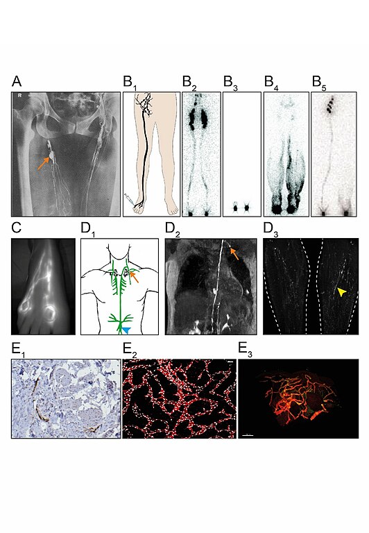
English: (A) Direct contrast X-ray lymphography involves the injection of an x-ray contrast agent e.g. Lipiodol. First, lymphatic vessels are identified by subcutaneous injection of a vital dye e.g. patent blue. Under local anesthetic, an incision is then made to expose the lymphatic vessel following the insertion of a needle into its lumen. The contrast agent can then be injected, and radiographs taken. Clear definition of lymphatic collectors and lymph nodes (arrow) can be achieved, but the procedure is invasive and rarely performed. (B1) Lymphoscintigraphy involves the injection of Technetium-99 into the web spaces between the toes (or fingers). Images using a gamma camera can be taken at specified time intervals and reveal lymph drainage channels with uptake of the contrast agent in regional lymph nodes. Removal of the contrast agent from the injection site and uptake in nodes reflect lymph transport (quantitative lymphoscintigraphy). (B2) Anterior view of a normal subject. (B3) Milroy patient carrying a VEGFR3 mutation showing no uptake by the initial lymphatics nor transport to the lymph nodes (functional aplasia). (B4) Lymphedema distichiasis syndrome patient carrying a FOXC2 mutation showing lymph reflux with dermal backflow seen as dark shading in the calf. (B5) Emberger patient carrying a GATA2 mutation exhibiting unilateral uptake, with the left leg showing no migration of tracer within the collecting lymphatics. (C) Indocyanine Green lymphography (ICGL) imaging of the right dorsal foot. Two indocyanine green intradermal injections are given in the toe web spaces followed by an immediate absorption of ICG into initial lymphatics. Excitation of the area of interest with laser or LED will emit fluorescence through the skin, that allows for real-time imaging with a near-infrared detector camera to visualize lymph drainage up the leg. Contractility of lymphatic collectors can be seen in real time. (D1+2) Dynamic contrast-enhanced magnetic resonance lymphangiogram (DCMRL) can image lymphatic vessels. Following bilateral injection of contrast under ultrasound guidance directly into an inguinal lymph node, central conducting vessels can be seen, like the cisterna chyli (arrowhead) and thoracic duct, highlighted in green in the drawing of the upper chest. The image is a T1 weighted MRI in which areas of contrast uptake appear bright demonstrating the thoracic duct terminating at the junction of the left subclavian and internal jugular veins, into which it drains (arrow). (D3) T2 weighted images demonstrate high signal in areas of static or slow-moving fluid. Vessel-like structures can be observed bilaterally in the legs (arrowhead). Whether these structures represent lymphatic or venous vessels remain controversial. Imaging of skin biopsies has advanced considerably with the development of modern microscopy techniques and the use of antibodies against lymphatic-specific markers. (E1) Immunohistochemistry (IHC) on paraffin-embedded 2D human skin sections showing podoplanin (PDPN) positive lymphatic vessels (brown DAB staining). Image taken at 20X magnification with a light microscope. (E2) Dermal lymphatic vasculature in the developing mouse embryo. Skin whole-mount preparation of E14.5 wildtype mouse embryos are visualized in 3D using a confocal microscope. Lymphatic endothelial cells are identified by Prox1 (white) and Vegfr3 (red) expression. Maximum intensity projections are shown. Scale bar = 100 µm. (E3). Whole-mount 3D human skin biopsy from a healthy control optically sectioned using a light sheet microscope and VIPAR analysis (164). Lymphatic endothelial cells are identified by PROX1 (green) and PDPN (red) expression. Scale bar = 100 µm. |
| Fecha | |
| Fuente | Trabajo propio |
| Autor | SGUL lymres |
Dr Malou van Zanten, St. George’s University of London; Dr Lakshmi Ratnam, St. Georges Hospital NHS Trust, London; Dr Rene Hägerling, Charité, Berlin; Sif Nielsen and eLearning Unit members Sheetal Kavia and Dhillon Khetani from St George’s, University of London (SGUL) have assisted with figure preparation.
Licencia
Yo, el titular de los derechos de autor de esta obra, la publico en los términos de la siguiente licencia:
Este archivo está disponible bajo la licencia Creative Commons Attribution-Share Alike 4.0 International.
- Eres libre:
- de compartir – de copiar, distribuir y transmitir el trabajo
- de remezclar – de adaptar el trabajo
- Bajo las siguientes condiciones:
- atribución – Debes otorgar el crédito correspondiente, proporcionar un enlace a la licencia e indicar si realizaste algún cambio. Puedes hacerlo de cualquier manera razonable pero no de manera que sugiera que el licenciante te respalda a ti o al uso que hagas del trabajo.
- compartir igual – En caso de mezclar, transformar o modificar este trabajo, deberás distribuir el trabajo resultante bajo la misma licencia o una compatible como el original.
Historial del archivo
Haz clic sobre una fecha y hora para ver el archivo tal como apareció en ese momento.
| Fecha y hora | Miniatura | Dimensiones | Usuario | Comentario | |
|---|---|---|---|---|---|
| actual | 11:48 29 ene 2021 | | 2250 × 3250 (754 kB) | SGUL lymres | Uploaded own work with UploadWizard |
Usos del archivo
La siguiente página usa este archivo:
Uso global del archivo
Las wikis siguientes utilizan este archivo:
- Uso en it.wikipedia.org