
Archivo:Nucleosome core particle 1EQZ v.5.jpg
Nucleosome_core_particle_1EQZ_v.5.jpg (620 × 560 píxeles; tamaño de archivo: 99 kB; tipo MIME: image/jpeg)
Resumen
| Descripción |
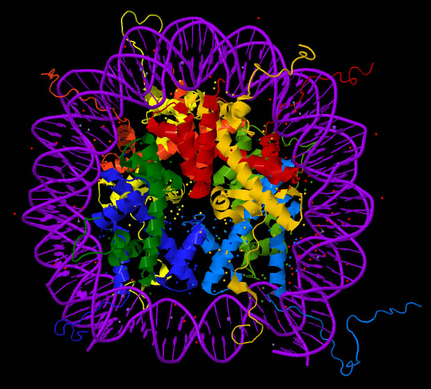
English: Nucleosome core particle, crystal structure (PDB ID: 1EQZ). Histones H2A, H2B, H3 and H4 are coloured. This image was created with Jmol (an open-source Java viewer for chemical structures in 3D http://www.jmol.org/) and Adobe Photoshop Elements. |
| Fecha | |
| Fuente |
Trabajo propio utilizando: the Protein Data Bank (PDB) structural data: |
| Autor | Darekk2 using the cited above Protein Data Bank (PDB) structural data |
Licencia
Attribution: The author of the work, the original authors of the Protein Data Bank (PDB) structural data and the molecular graphics program used. PDB data and software citation example:
Image of PDB (Protein Data Bank) ID 1EQZ (Harp, J.M., Hanson, B.L., Timm, D.E., Bunick, G.J. (2000) Asymmetries in the nucleosome core particle at 2.5 A resolution. Acta Crystallogr., Sect.D 56: 1513-1534) created with Jmol (an open-source Java viewer for chemical structures in 3D. http://www.jmol.org/).
The RCSB Protein Data Bank (PDB) states on its website in the Policies & References section:
http://www.rcsb.org/pdb/static.do?p=general_information/about_pdb/policies_references.html archive copy at the Wayback Machine
Data files contained in the PDB archive (ftp://ftp.wwpdb.org) are free of all copyright restrictions and made fully and freely available for both non-commercial and commercial use. Users of the data should attribute the original authors of that structural data. (...) Images created using PDB data and other software should cite the PDB ID and the molecular graphics program used.
the original authors of the Protein Data Bank (PDB) structural data
and the molecular graphics program used
- Eres libre:
- de compartir – de copiar, distribuir y transmitir el trabajo
- de remezclar – de adaptar el trabajo
- Bajo las siguientes condiciones:
- atribución – Debes otorgar el crédito correspondiente, proporcionar un enlace a la licencia e indicar si realizaste algún cambio. Puedes hacerlo de cualquier manera razonable pero no de manera que sugiera que el licenciante te respalda a ti o al uso que hagas del trabajo.
- compartir igual – En caso de mezclar, transformar o modificar este trabajo, deberás distribuir el trabajo resultante bajo la misma licencia o una compatible como el original.
Historial del archivo
Haz clic sobre una fecha y hora para ver el archivo tal como apareció en ese momento.
| Fecha y hora | Miniatura | Dimensiones | Usuario | Comentario | |
|---|---|---|---|---|---|
| actual | 22:16 9 oct 2012 | | 620 × 560 (99 kB) | Darekk2 | changed colors and rotated 180 deg |
| 12:25 9 oct 2012 |  | 596 × 567 (94 kB) | Darekk2 | User created page with UploadWizard |
Usos del archivo
Las siguientes páginas usan este archivo:
Uso global del archivo
Las wikis siguientes utilizan este archivo:
- Uso en ar.wikipedia.org
- Uso en bs.wikipedia.org
- Uso en ca.wikipedia.org
- Uso en el.wikipedia.org
- Uso en en.wikipedia.org
- Uso en ja.wikipedia.org
- Uso en vi.wikipedia.org